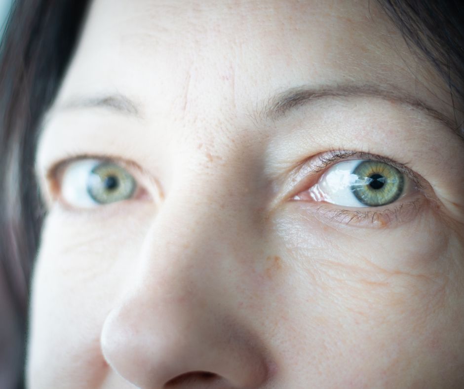
27 Mar Exophthalmia VS. Enophthalmia: Understanding Bulging And Sunken Eyes
Our eyes can reveal a lot about our health, but what happens when they start to look noticeably different? Exophthalmia (bulging eyes) and enophthalmia (sunken eyes) are two conditions that affect the position of the eyes within their sockets, often signaling underlying medical concerns. In this blog, we’ll break down the differences between exophthalmia and enophthalmia, their causes, symptoms, and potential treatment options, so you can understand more about your eyes.
What Is Exophthalmia
Exophthalmia, also called exophthalmos, refers to a condition where one or both eyes appear to protrude out of the eye socket. When the muscles and fatty tissues behind the eye become inflamed, they can press on the eye. This swelling can cause pain and other ocular symptoms.
It has the potential to cause functional vision problems, in addition to being an aesthetic concern. In all but the rarest cases, it is linked to underlying health problems, making an understanding of what causes it an important first step.
What Is Enophthalmia
Enophthalmia is characterized by a sinking of the eyeball into the eye socket, giving the eye a severe sunken appearance. This occurs due to an underdevelopment of the structures encasing the eye, generally due to trauma or other complications. The most common cause is a medial orbital floor fracture.
The severity varies based on how far the orbital rim has dislocated. Research suggests that even a small 7-mm shift in the lateral wall of the eye socket can increase its volume by about 16%. This significant change can have a major impact on the position and function of the eye.
Differences Between Exophthalmia And Enophthalmia
Exophthalmia and enophthalmia are two opposite clinical entities that influence the position of the eyeball in the eye socket. Specialists frequently rely on exact metrics to differentiate the two states.
Exophthalmia, or proptosis, occurs when the eye protrudes forward. Conversely, enophthalmia is characterized by the eye sinking more deeply into the orbital cavity. A measurable difference greater than 2 millimeters (mm) between the two eyes is diagnostic in both conditions.
For exophthalmia, an Anterior Globe Projection (AGP) more than 20 mm, as measured using a Hertel exophthalmometer, is characteristic. This instrument has the lateral orbital rim as a tactile reference point for precision.
Enophthalmia is typically acknowledged when the AGP is below 10 mm. Enophthalmos is clinically evident once the AGP is less than 12 mm. A difference of more than 3 mm between the two eyes may be an important diagnostic characteristic of the disease as well.
Treatment For Exophthalmia
Treatment for exophthalmia depends on the severity of the condition and requires a personalized approach. The primary goal is to manage symptoms and address the underlying cause to prevent further progression.
Initial treatment focuses on symptom relief, including the use of artificial tears or ointments to alleviate dryness and irritation. Patients experiencing light sensitivity may benefit from specialized sunglasses. Lifestyle modifications, such as quitting smoking, can also help reduce inflammation and improve overall eye health.
In more severe cases, medical interventions may be necessary. Corticosteroids or other anti-inflammatory medications can help reduce swelling, while orbital decompression surgery may be required to create more space within the eye socket and restore proper positioning of the eye. In some cases, eye muscle surgery or eyelid procedures can help improve both function and appearance.
Regular monitoring by an eye doctor is essential to track any changes and adjust treatment as needed. Exophthalmia often stabilizes over time, but proactive management can significantly improve long-term outcomes.
Treatment For Enophthalmia
Treatment for enophthalmia depends on its underlying cause and typically requires a multidisciplinary approach. In cases of traumatic enophthalmia, orbital fractures may lead to the eye sinking further into the socket. Surgical intervention is often necessary to repair fractures, reposition displaced bones, and restore the natural anatomy of the orbit.
Age-related enophthalmia occurs due to fat atrophy around the eye, leading to a sunken appearance. This condition is often managed with non-surgical options such as dermal fillers or fat grafting to restore lost volume and improve symmetry.
In cases where enophthalmia results from tumours or inflammatory orbital diseases, treatment depends on the specific diagnosis. Management may involve surgery, radiation therapy, or medical treatment to address the underlying condition and prevent further progression.
A comprehensive evaluation by an optometrist is essential to determine the most effective treatment plan based on the individual’s condition.
Conclusion
We know too well the importance of maintaining eye health. Exophthalmia and enophthalmia are eye conditions that can be intimidating, but knowing what causes them and what treatment options are available helps immensely. By identifying the symptoms sooner rather than later, you can prevent vision loss and increase your overall quality of life. Regardless of the treatment you choose, treatments are most effective when customized to fit individual needs, so being educated and working with a trusted eye doctor is crucial.
By getting regular checkups and educating yourself about the disease, you can stay one step ahead of dangerous complications. If you experience new symptoms or changes to the appearance of your eyes, don’t hesitate to reach out to Dr. D’Orio Eyecare today. Visit https://drdorioeyecare.com/book-appointment/ or call us at 416-656-2020 for our Toronto location, or 416-661-5555 for our North York location.


