WE USE THE NEWEST EQUIPMENT
With improved technology healthcare professionals are able to provide patients with improved care and comfort. These new innovations have helped to change and improve the way an optometrist can manage or treat certain eye diseases. The OCT and Retinal Camera has allowed optometrists to diagnose and detect early signs of treatable eye conditions. This technology has allowed for better outcomes for disease treatment because of early detection and increased specificity.
Having the newest technology is essential in diagnosing and managing many types of eye diseases. Certain eye conditions prognosis is dictated by early detection and treatment. These new innovations will help detect and diagnosis certain eye conditions so the best treatment outcome can be provided towards each one of our patients.
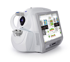
Cirrus 500 Optical Coherence Tomography
With improved technology healthcare professionals are able to provide patients with improved care and comfort. These new innovations have helped to change and improve the way an optometrist can manage or treat certain eye diseases. The OCT and Retinal Camera has allowed optometrists to diagnose and detect early signs of treatable eye conditions. This technology has allowed for better outcomes for disease treatment because of early detection and increased specificity.
Having the newest technology is essential in diagnosing and managing many types of eye diseases. Certain eye conditions prognosis is dictated by early detection and treatment. These new innovations will help detect and diagnosis certain eye conditions so the best treatment outcome can be provided towards each one of our patients.
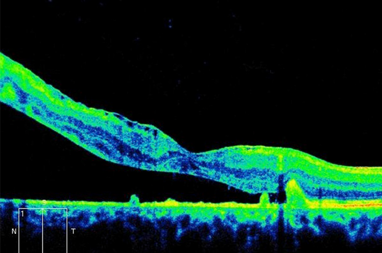
Retinal Detachment (SEROUS)
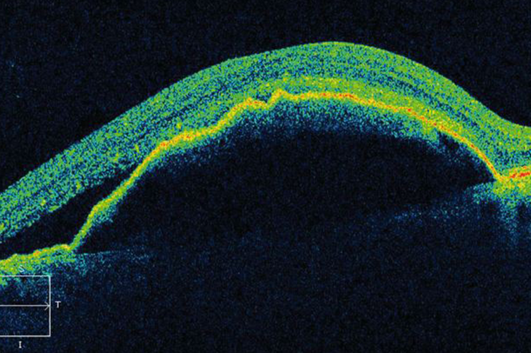
PIGMENT Epithelial detachment
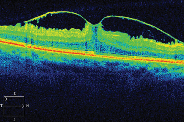
VITREOMACULAR TRACTION
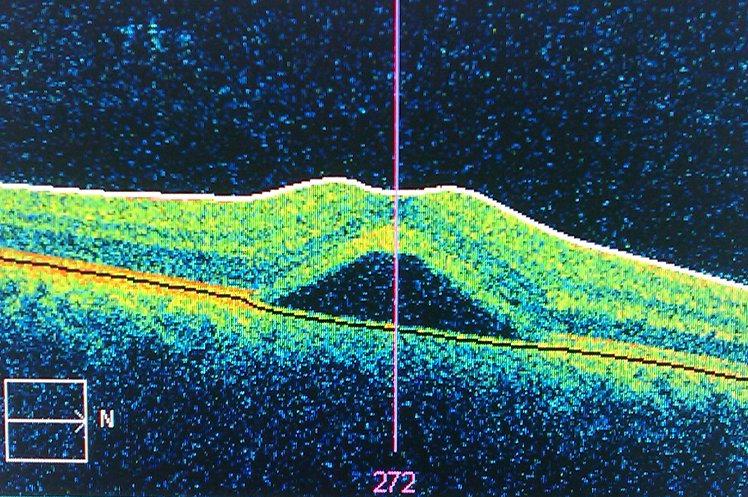
CENTRAL SEROUS CHORIORETINOPATHY
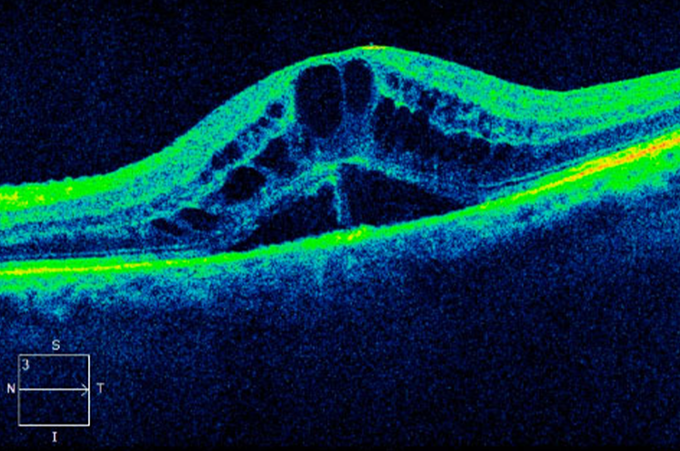
DIABETIC MACULAR EDEMA
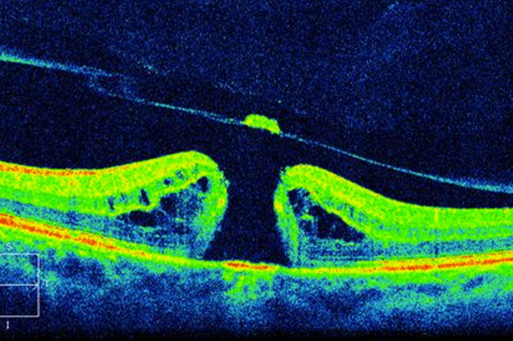
MACULAR HOLE
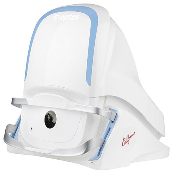
OPTOS CALIFORNIA CAMERA
The biggest advantage of the Optos California camera is that it has the ability to capture the largest field of view possible in one image. Our eye care professionals are now able to see 50% more of the retina when compared to other conventional imaging devices.This allows our doctors to proactively detect and diagnose our patients more efficiently.
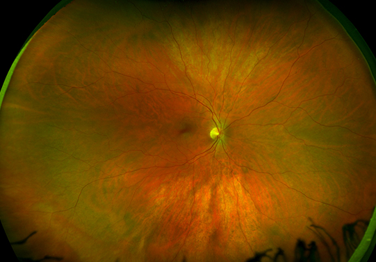
healthy retina
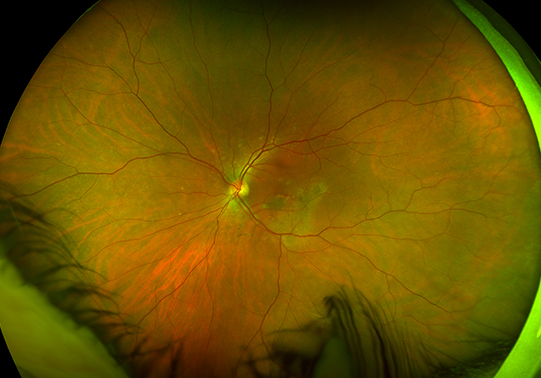
age-related macular degeneration (amd)
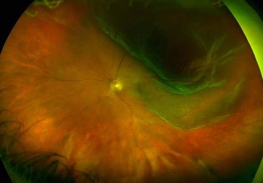
Retinal detachment
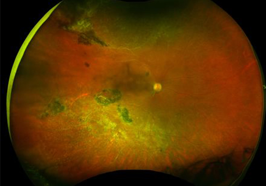
retinoschisis
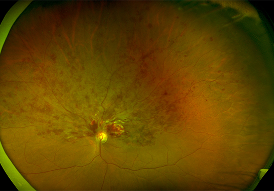
retinal vein occlusion
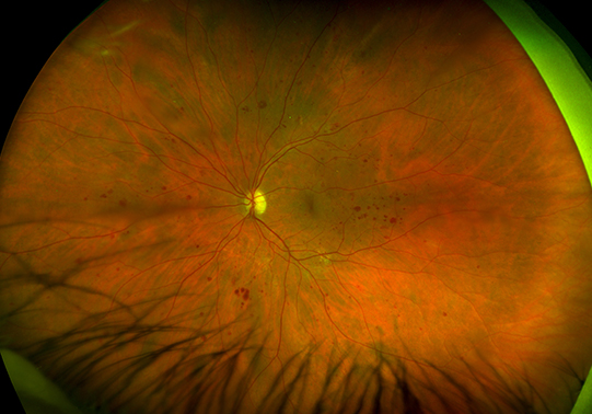
diabetic retinopathy
Other Medical Equipment We Use
NIDEK ARK-530A AUTOREFRACTOR
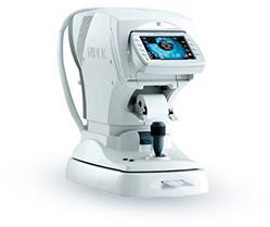
The combination of new measuring principle – Pupil Zone Imaging Method – and unique technology – SLD – offers high accuracy and reliability in refraction measurement.
The NIDEK ARK-530A also adopts the advanced Pupil Zone Imaging Method for refraction measurement, which analyzes a wider area (Max. ø4 mm) to obtain more reliable and realistic data that is closer to subjective refraction.
HUMPHREY VISUAL FIELD
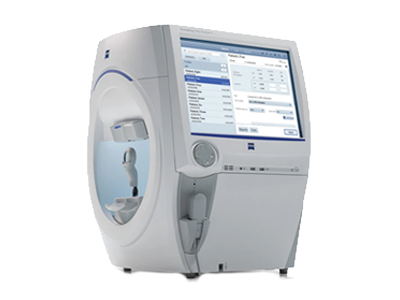
Providing advanced analysis with comprehensive connectivity options validated by more than 30 years of research, design and clinical experience, the Humphrey Field Analyzer (HFA) is the accepted standard of care in glaucoma diagnosis and management. With over 65,000 installed units worldwide, the HFA is the premier automated visual field analyzer. A visual field test is an eye examination that can detect dysfunction in central and peripheral vision.
Additionally, the Humphrey Visual Field (HVF) is a special automated procedure used to measure the entire area of peripheral vision that can be seen when an eye is focused on a central point. This machine assists in diagnosing and monitoring neurological conditions such as pituitary tumors, optic neuropathies, strokes and more.
BLEPHEX
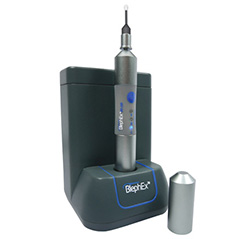
BlephEx is a revolutionary new patented hand piece, used to very precisely and carefully, spin a medical grade micro-sponge along the edge of your eyelids and lashes, removing scurf and debris and exfoliating your eyelids.
BlephEx is a very useful addition to the lid hygiene / dry eye armamentarium. The device effectively clears debris from the lashes and lid margins and to my surprise contributes to down-regulating inflammation along with a subjective improvement in symptoms.
INFLAMMADRY
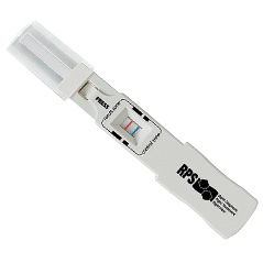
InflammaDry is the first and only, rapid result, in-office test that detects elevated levels of MMP-9, an inflammatory marker that is consistently elevated in the tears of patients with dry eye disease. All other dry eye tests measure tear production and stability.
Using a simple 4-step process, InflammaDry recognizes elevated levels of MMP-9, to identify patients that may otherwise be missed with other dry eye testing methods.
TEARLAB
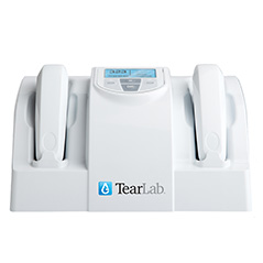
TearLab is an objective and quantitative point-of-care diagnostic test that provides precise and predictive information.
The TearLab Osmolarity System* is intended to measure the osmolarity of human tears to aid in the diagnosis of dry eye disease in patients suspected of having dry eye disease, in conjunction with other methods of clinical evaluation.
*TearLab is for professional in vitro diagnostic use only.
ANTARES S40PTIK
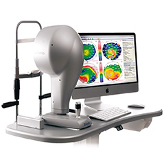
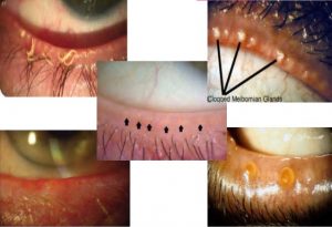
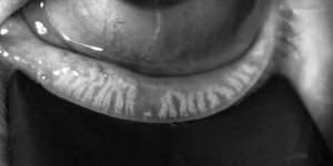
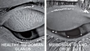
ANTARES new corneal topographer with advanced tear-film analysis. Boasts a new high resolution digital colour camera and advanced algorithms, to bring you user friendly, full featured Corneal Topographic data, TBUT analysis, as well as MGD analysis.
Features:
- Corneal Topography
- Contact lens fitting with Autofit
- Keratoconus screening with classification
- Dynamic view of non-invasive break-up time
- Tear Meniscus Height Imaging and Measurement
- Interference Pattern Imaging of Lipid Layer
- Imaging of the Tear Film Dynamics (Video)
- Fluorescein Imaging
- Meibomian Glands Imaging and Analysis
ILUX
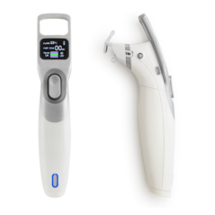
The iLux is a handheld portable device that is used for treating dry eye disease. It treats dry eye disease that is associated with meibomian gland dysfunction by using light-based heat and compression, under direct visualization by the physician under a magnifying lens. The sterile, single-use, iLux Smart Tip has an inner pad that slips behind the eyelid being treated and an outer pad that’s pressed against the outer surface of the eyelid during heating and compression; the tip contains temperature sensors that maintain safe, therapeutic heat levels during treatment. This treatment help by opening or unclogging the meibomian gland orfices so oil can be expressed passively after every blink.
E > EYE
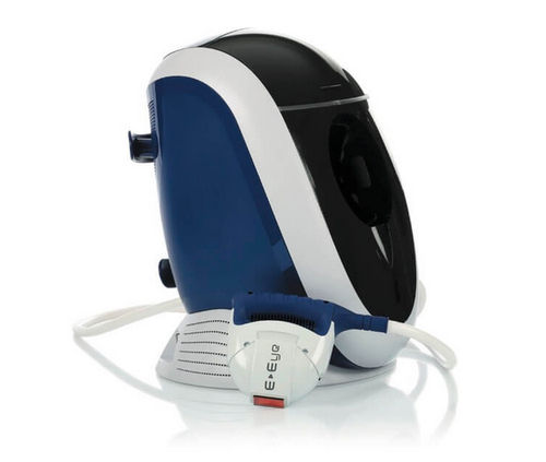
The E-Eye is a device that generates a new type of polychromatic pulsed light by producing perfectly calibrated and homogeneously sequenced light pulses. The sculpted pulses are delivered under the shape of regulated train pulses. The energy, spectrum and time period are precisely set to stimulate the meibomian glands to improve their function. The proposed mechanism of action of this is neurological. Localized treatment is directed at the lower eyelid and the lower arcades of the parasympathetic nerve. Infrared nerve stimulation induces a “return to normal” activity of the meibomian glands. Treatment improves meibomian gland function and reduces the inflammatory cascades.


